

MedFriendly®


Biopsy
Biopsy has the following meanings in the field of
medicine:
1. The process of removing living tissue or cells from
organs or other body parts of patients for examination
under a microscope or in a culture to help make a
diagnosis, follow the course of a disease, or estimate a
prognosis. A culture is an artificial way to grow cells or
tissues in the laboratory. Most biopsies are minor
procedures and do not require the area where the
sample is being taken from to be numbed with
medication. Some biopsies are more serious and/or
complicated and require that the appropriate body part
be numbed.
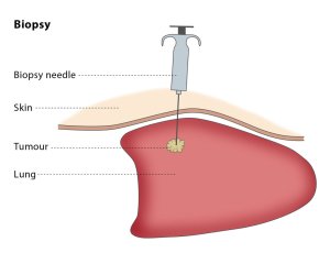
A lung biopsy (needle biopsy).
FEATURED BOOK: Anticancer: A New Way of Life
2. A sample obtained by using the above procedure. This definition of biopsy is more
accurately known as a "biopsy specimen."
WHY IS A BIOPSY PERFORMED?
A biopsy is performed because it is generally an accurate technique that can help
diagnose many illnesses, such as cancer. Cancer is any of a large group of malignant
diseases characterized by an abnormal, uncontrolled growth of new cells in one of the
body organs or tissues. When describing cancer cells, malignant means that the newly
formed tissues are made of abnormally structured and primitive-looking cells that grow
uncontrollably, spread throughout the body, and invade surrounding tissues.
"Where Medical Information is Easy to Understand"™
Biopsies can help establish if tumors are malignant or benign.
Tumors are abnormal masses of tissue that form when cells in a
certain area of the body reproduce at an increased rate. Unlike
malignant tumors, benign tumors stay in a localized area of the
body. Malignant tumors are cancerous whereas benign tumors are
not. When samples of tumors are examined under the microscope,
malignant tumors have many characteristics that help clearly tell
them apart from benign tumors. Since malignant tumors have the
capability of spreading, biopsies of tissues and lymph nodes that
surround a tumor are often performed to determine if the cancer has
spread. Lymph nodes are small egg shaped structures found
throughout the body that help fight against infection.
Biopsies are also useful in helping to determine the cause of infections or inflammation that has gone
unexplained. Some biopsies are done to obtain samples of healthy tissue so that doctors can tell if the
tissue can be transplanted into someone else's body who needs it. More often than not, however,
unhealthy tissue is biopsied as opposed to healthy tissue.
ARE THERE DIFFERENT TYPES OF BIOPSIES?
Yes, there are many different types of biopsies, some of which will be described here:
SKIN/MUSCLE BIOPSY: In this type of biopsy, a small piece of skin or muscle is cut away from the body
and sent to be analyzed in the laboratory. This procedure usually only takes a few seconds to do after the
area of the body where the sample is being taken from has been numbed. Stitches are sometimes used to
sew the skin back together. In small biopsies, the skin may heal fine without stitches.
NEEDLE BIOPSY: In this type of biopsy, a needle is inserted through the skin and into the organ that is
believed to be abnormal is some way. At the end of the needle, a cutting device may be fitted on the tip to
help remove a piece of the tissue for examination. A small, sharp pinch is usually felt at the site of the
biopsy.
The doctor usually uses the assistance of tests that can take pictures of the inside of the body while the
biopsy is being performed so he/she knows where to direct the needle. This technique is known as guided
biopsy and it is more safe and accurate than doing a biopsy without the guided help. Tests that are often
used to help the doctor in guided biopsies are ultrasounds and CT scans. An ultrasound is a procedure
that uses types of sound waves to produce images of the body. CT (computerized tomography) scanning
is an advanced imaging technique that uses x-rays and computer technology to produce more clear and
detailed pictures than a traditional x-ray.
The needles used in needle biopsies can be large or very small. Very thin and small needles have helped
doctors take tissue samples from areas of the body (such as the glands that produce saliva) that were
dangerous to take from with large needles. A needle biopsy is also known as a percutaneous biopsy.
ASPIRATION BIOPSY: This is a type of needle biopsy (see above) in which the needle is placed into a
tumor or abnormal mass to remove cells. Most aspiration biopsies only require that the specific body part
being examined be numbed. Sometimes, however, general anesthesia is required. General anesthesia is
loss of sensation and consciousness in the entire body that is caused by administering certain
medications. This type of biopsy is also known as a needle aspiration biopsy.
ENDOSCOPIC BIOPSY: In this type of biopsy, a flexible tube with a viewing lens (known as an
endoscope) is passed through a tiny cut in the skin and then into the organ that needs to be examined. An
attachment is passed through the endoscope to take a sample of the area of concern. The attachment
used to remove tissues is known as forceps. Brushes are the attachments used to remove cells.
EXCISIONAL BIOPSY: In this type of biopsy, the surgeon removes the entire area of concern (such as a
lumpy mass) as opposed to only removing a part of it. The entire area that is removed will be sent to the
laboratory for analysis. For lumps or masses located in the breast that were detected with a
mammography, the surgeon may first need to inject a die or thin wires to identify the abnormal area. A
mammography is an X-ray procedure that is performed as a screening test for women who want to detect
breast cancer at an early stage.
OPEN BIOPSY: This type of biopsy is part of an operation in which an area of the body (such as the
chest or the belly) needs to be cut open to view a diseased area so that a tissue sample can be obtained.
The surgery is typically performed under general anesthesia. The surgeon can send the tissue sample to
the laboratory for rapid analysis, which helps make the decision as to whether the entire area needs to be
removed. An open biopsy is performed when an endoscopic biopsy is not possible or when it is likely that
the diseased organ or tissue will need to be removed. An endoscopic biopsy is sometimes called a closed
biopsy because it only requires a tiny cut to be made in the skin.
HOW ARE THE RESULTS OF BIOPSIES OBTAINED?
When a biopsy is performed, the sample is sent to the laboratory for analysis. If the situation is urgent,
biopsy results can be ready within minutes. An example of an urgent situation would be a patient that has
had a body part opened that may have cancer in it. It would be urgent to find out if the tissue sample is
cancerous so that the diseased area can be removed immediately. In less serious situations, it may take
a few days for biopsy results to come back.
A special stain is placed on the sample of tissues or cells that helps highlight certain areas that can be
seen under a microscope. The samples are frozen or smeared on a slide that is then examined under a
microscope. Normal and abnormal cells have certain appearances that can be detected under the
microscope. The presence and type of abnormal features in cells and tissues is what helps lead to a
diagnosis. The sample may also be embedded in wax so that it is given a firm consistency, can better be
sliced into very thin slices, placed on a slide, and examined. The wax procedure is more time consuming,
taking about 24 hours.
If fluid samples were taken that contain cells, the fluid is sometimes spun at high speeds in a device called
a centrifuge. Spinning the fluid at high speeds helps to pack together cells inside the fluid. The fluid is then
placed on a small slide for examination. The cells are preserved on the slide and stains are applied to
better view them under the microscope.
A cytologist examines the cells for abnormalities, paying special attention to the size, shape, and structure
on the nucleus (center of the cell). A cytologist is someone who specializes in cytology, an area of biology
that deals with the study of the structure, function, multiplication, life history, problems, diseases, and
chemistry of the cell.
As was mentioned earlier, some biopsies are cultured, meaning that the sample is placed in a special
laboratory container to see if it grows. Sometimes, specific antibodies are introduced to the tissue sample
to see how it reacts. Antibodies are types of proteins that are formed by the body to destroy foreign
proteins known as antigens. When an antibody meets an antigen, inflammation results. Cultures and
antibody tests are used when the doctor is examining cases of infection or inflammation.
In some kidney biopsies, an extremely powerful microscope (known as an electron microscope) is used to
help determine the type of cell that makes up the tumor. The kidneys are two organs located on each side
of the spine, behind the stomach. The kidneys filter (remove) wastes from the blood. There are other
specialized techniques used to analyze the sample of tissue or cells. These specialized techniques uses
special stains, antibodies, and enzymes. An enzyme is a type of protein that helps produce chemical
reactions in the body. These specialized procedures take longer to do but are generally more accurate
than non-specialized procedures regarding diagnosis and prognosis.
WHAT ARE THE RISKS INVOLVED IN A BIOPSY?
The risks involved in a biopsy include pain, bleeding, and infection.
WHAT IS THE ORIGIN OF THE TERM, BIOPSY?
Biopsy comes from the Greek word "bios" meaning "life," and the Greek word "opsis" meaning "vision." Put
the words together, and you have "life vision."